Showing posts sorted by relevance for query malocclusion. Sort by date Show all posts

Tuesday, November 17, 2009

Malocclusion: Disease of Civilization, Part VI

Early Postnatal Face and Jaw Development

The face and jaws change more from birth to age four than at any other period of development after birth. At birth, infants have no teeth and their skull bones have not yet fused, allowing rapid growth. This period has a strong influence on the development of the jaws and face. The majority of malocclusions are established by the end this stage of development. Birth is the point at which the infant begins using its jaws and facial musculature in earnest.

The development of the jaws and face is very plastic, particularly during this period. Genes do not determine the absolute size or shape of any body structure. Genes carry the blueprint for all structures, and influence their size and shape, but structures develop relative to one another and in response to the forces applied to them during growth. This is how orthodontists can change tooth alignment and occlusion by applying force to the teeth and jaws.

Influences on Early Postnatal Face and Jaw Development

In 1987, Miriam H. Labbok and colleagues published a subset of the results of the National Health Interview survey (now called NHANES) in the American Journal of Preventive Medicine. Their article was provocatively titled "Does Breast-feeding Protect Against Malocclusion"? The study examined the occlusion of nearly 10,000 children, and interviewed the parents to determine the duration of breast feeding. Here's what they found:

The longer the infants were breastfed, the lower their likelihood of major malocclusion. The longest category was "greater than 12 months", in which the prevalence of malocclusion was less than half that of infants who were breastfed for three months or less. Hunter-gatherers and other non-industrial populations typically breastfeed for 2-4 years, but this is rare in affluent nations. Only two percent of the mothers in this study breastfed for longer than one year.

The prevalence and duration of breastfeeding have increased dramatically in the US since the 1970s, with the prevalence doubling between 1970 and 1980 (NHANES). The prevalence of malocclusion in the US has decreased somewhat in the last half-century, but is still very common (NHANES).

Several, but not all studies have found that infants who were breastfed have a smaller risk of malocclusion later in life (1, 2, 3). However, what has been more consistent is the association between non-nutritive sucking and malocclusion. Non-nutritive sucking (NNS) is when a child sucks on an object without getting calories out of it. This includes pacifier sucking, which is strongly associated with malocclusion*, and finger sucking, which is also associated to a lesser degree.

The longer a child engages in NNS, the higher his or her risk of malocclusion. The following graph is based on data from a study of nearly 700 children in Iowa (free full text). It charts the prevalence of three types of malocclusion (anterior open bite, posterior crossbite and excessive overjet) broken down by the duration of the NNS habit:

As you can see, there's a massive association. Children who sucked pacifiers or their fingers for more than four years had a 71 percent chance of having one of these three specific types of malocclusion, compared with 14 percent of children who sucked for less than a year. The association between NNS and malocclusion appeared after two years of NNS. Other studies have come to similar conclusions, including a 2006 literature review (1, 2, 3).

Bottle feeding, as opposed to direct breast feeding, is also associated with a higher risk of malocclusion (1, 2). One of the most important functions of breast feeding may be to displace NNS and bottle feeding. Hunter-gatherers and non-industrial cultures breast fed their children on demand, typically for 2-4 years, in addition to giving them solid food.

In my opinion, it's likely that NNS beyond two years of age, and bottle feeding to a lesser extent, cause a large proportion of the malocclusions in modern societies. Pacifier use seems to be particularly problematic, and finger sucking to a lesser degree.

How Do Breastfeeding, Bottle Feeding and NNS Affect Occlusion?

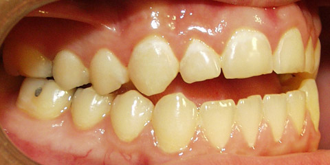
Since jaw development is influenced by the forces applied to them, it makes sense that the type of feeding during this period could have a major impact on occlusion. Children who have a prolonged pacifier habit are at high risk for open bite, a type of malocclusion in which the incisors don't come together when the jaws are closed. You can see a picture here. The teeth and jaws mold to the shape of the pacifier over time. This is because the growth patterns of bones respond to the forces that are applied to them. I suspect this is true for other parts of the skeleton as well.

Any force applied to the jaws that does not approximate the natural forces of breastfeeding or chewing and swallowing food, will put a child at risk of malocclusion during this period of his or her life. This includes NNS and bottle feeding. Pacifier sucking, finger sucking and bottle feeding promote patterns of muscular activity that result in weak jaw muscles and abnormal development of bony structures, whereas breastfeeding, chewing and swallowing strengthen jaw muscles and promote normal development (review article). This makes sense, because our species evolved in an environment where the breast and solid foods were the predominant objects that entered a child's mouth.

What Can We do About it?

In an ideal world (ideal for occlusion), mothers would breast feed on demand for 2-4 years, and introduce solid food about halfway through the first year, as our species has done since the beginning of time. For better or worse, we live in a different world than our ancestors, so this strategy will be difficult or impossible for many people. Are there any alternatives?

Parents like bottle feeding because it's convenient. Milk can be prepared in advance, the mother doesn't have to be present, feeding takes less time, and the parents can see exactly how much milk the child has consumed. One alternative to bottle feeding that's just as convenient is cup feeding. Cup feeding, as opposed to bottle feeding, promotes natural swallowing motions, which are important for correct development. The only study I found that examined the effect of cup feeding on occlusion found that cup-fed children developed fewer malocclusion and breathing problems than bottle-fed children.

Cup feeding has a long history of use. Several studies have found it to be safe and effective. It appears to be a good alternative to bottle feeding, that should not require any more time or effort.

What about pacifiers? Parents know that pacifiers make babies easier to manage, so they will be reluctant to give them up. Certain pacifier designs may be more detrimental than others. I came across the abstract of a study evaluating an "orthodontic pacifier" called the Dentistar, made by Novatex. The frequency of malocclusion was much lower in children who did not use a pacifier or used the Dentistar, than in those who used a more conventional pacifier. This study was funded by Novatex, but was conducted at Heinrich Heine University in Dusseldorf, Germany**. The pacifier has a spoon-like shape that allows normal tongue movement and exerts minimal pressure on the incisors. There may be other brands with a similar design.

The ideal is to avoid bottle feeding and pacifiers entirely. However, cup feeding and orthodontic pacifiers appear to be acceptable alternatives that minimize the risk of malocclusion during this critical developmental window.


* Particularly anterior open bite and posterior crossbite.

** I have no connection whatsoever to this company. I think the results of the trial are probably valid, but should be replicated.

Saturday, October 3, 2009

Malocclusion: Disease of Civilization, Part II

The Nature of the Problem

In 1973, the US Centers for Disease Control and Prevention (CDC) published the results of a National Health Survey in which it examined the dental health of American youths nationwide. The following description was published in a special issue of the journal Pediatric Dentistry (1):
The 1973 National Health Survey reported 75% of children, ages 6 to 11 years, and 89% of youths, ages 12 to 17 years, have some degree of occlusal disharmony [malocclusion]; 8.7% of children and 13% of youth had what was considered a severe handicapping malocclusion for which treatment was highly desirable and 5.5% of children and 16% of youth had a severe handicapping malocclusion that required mandatory treatment.
89% of youths had some degree of malocclusion, and 29% had a severe handicapping malocclusion for which treatment was either highly desirable or mandatory. Fortunately, many of these received orthodontics so the malocclusion didn't persist into adulthood.

This is consistent with another survey conducted in 1977, in which 38% of American youths showed definite or severe malocclusion. 46% had occlusion that the authors deemed "ideal or acceptable" (2).

The trend continues. The CDC National Health and Nutrition Examination Survey III (NHANES III) found in 1988-1991 that approximately three fourths of Americans age 12 to 50 years had some degree of malocclusion (3).

The same holds true for Caucasian-Americans, African-Americans and Native Americans in the US, as well as other industrial nations around the world. Typically, only 1/3 to 1/2 of the population shows good (but not necessarily perfect) occlusion (4- 8).

In the next post, I'll review some of the data from non-industrial and transitioning populations.


Malocclusion: Disease of Civilization


1. Pediatr. Dent. 17(6):1-6. 1995-1996
2. USPHS Vital and Health Statistics Ser. 11, no 162. 1977
3. J. Dent. Res. Special issue. 75:706. 1996. Pubmed link.
4. The Evaluation of Canadian Dental Health. 1959. Describes Canadian occlusion.
5. The Effects of Inbreeding on Japanese Children. 1965. Contains data on Japanese occlusion.
6. J. Dent. Res. 35:115. 1956. Contains data on both industrial and non-industrial cultures (Pukapuka, Fiji, New Guinea, U.S.A. and New Zealand).
7. J. Dent. Res. 44:947. 1965 (
free full text). Contains data on Caucasian-Americans and African-Americans living in several U.S. regions, as well as data from two regions of Germany. Only includes data on Angle classifications, not other types of malocclusion such as crossbite and open bite (i.e., the data underestimate the total prevalence of malocclusion).
8. J. Dent. Res. 47:302. 1968 (free full text). Contains data on Chippewa Native Americans in the U.S., whose occlusion was particularly bad, especially when compared to previous generations.

Saturday, October 10, 2009

Malocclusion: Disease of Civilization, Part III

Normal Human Occlusion

In 1967, a team of geneticists and anthropologists published an extensive study of a population of Brazilian hunter-gatherers called the Xavante (1). They made a large number of physical measurements, including of the skull and jaws. Of 146 Xavante examined, 95% had "ideal" occlusion, while the 5% with malocclusion had nothing more than mild cro
wding of the incisors (front teeth). The authors wrote:
Characteristically, the Xavante adults exhibited broad dental arches, almost perfectly aligned teeth, end-to-end bite, and extensive dental attrition [tooth wear].
In the same paper, the author presents occlusion statistics for three other cultures. According to the papers he cites, in Japan, the prevalence of malocclusion was 59%, and in the US (Utah), it was 64%. He also mentions another native group living near the Xavante, part of the Bakairi tribe, living at a government post and presumably eating processed food. The prevalence of malocclusion was 45% in this group.

In 1998, Dr. Brian Palmer (DDS) published a paper describing some of the collections of historical skulls he had examined over the years (2):
...I reviewed an additional twenty prehistoric skulls, some dated at 70,000 years old and stored in the Anthropology Department at the University of Kansas. Those skulls also exhibited positive [good] occlusions, minimal decay, broad hard palates, and "U-shaped" arches.

The final evaluations were of 370 skulls preserved at the Smithsonian Institution in Washington, D.C. The skulls were those of prehistoric North American plains Indians and more contemporary American skulls dating from the 1920s to 1940s. The prehistoric skulls exhibited the same features as mentioned above, whereas a significant destruction and collapse of the oral cavity were evident in the collection of the more recent skulls. Many of these more recent skulls revealed severe periodontal disease, malocclusions, missing teeth, and some dentures. This was not the case in the skulls from the prehistoric periods...
The arch is the part of the upper jaw inside the "U" formed by the teeth. Narrow dental arches are a characteristic feature of malocclusion-prone societies. The importance of arch development is something that I'll be coming back to repeatedly. Dr. Palmer's paper includes the following example of prehistoric (L) and modern (R) arches:


Dr. Palmer used an extreme example of a modern arch to illustrate his point, however, arches of this width are not uncommon today. Milder forms of this narrowing affect the majority of the population in industrial nations.

In 1962, Dr. D.H. Goose published a
study of 403 British skulls from four historical periods: Romano-British, Saxon, medieval and modern (3). He found that the arches of modern skulls were less broad than at any previous time in history. This followed an earlier study showing that modern British skulls had more frequent malocclusion than historical skulls (4). Goose stated that:
Although irregularities of the teeth can occur in earlier populations, for example in the Saxon skulls studied by Smyth (1934), the narrowing of the palate seems to have occurred in too short a period to be an evolutionary change. Hooton (1946) thinks it is a speeding up of an already long standing change under conditions of city life.
Dr. Robert Corruccini published several papers documenting narrowed arches in one generation of dietary change, or in genetically similar populations living rural or urban lifestyles (reviewed in reference #5). One was a st
udy of Caucasians in Kentucky, in which a change from a traditional subsistence diet to modern industrial food habits accompanied a marked narrowing of arches and increase in malocclusion in one generation. Another study examined older and younger generations of Pima Native Americans, which again showed a reduction in arch width in one generation. A third compared rural and urban Indians living in the vicinity of Chandigarh, showing marked differences in arch breadth and the prevalence of malocclusion between the two genetically similar populations. Corruccini states:
In Chandigarh, processed food predominates, while in the country coarse millet and locally grown vegetables are staples. Raw sugar cane is widely chewed for enjoyment rurally [interestingly, the rural group had the lowest incidence of tooth decay], and in the country dental care is lacking, being replaced by chewing on acacia boughs which clean the teeth and are considered medicinal.
Dr. Weston Price came to the same conclusion examining prehistoric skulls from South America, Australia and New Zealand, as well as their living counterparts throughout the world that had adhered to traditional cultures and foodways. From Nutrition and Physical Degeneration:
In a study of several hundred skulls taken from the burial mounds of southern Florida, the incidence of tooth decay was so low as to constitute an immunity of apparently one hundred per cent, since in several hundred skulls not a single tooth was found to have been attacked by tooth decay. Dental arch deformity and the typical change in facial form due to an inadequate nutrition were also completely absent, all dental arches having a form and interdental relationship [occlusion] such as to bring them into the classification of normal.
Price found that the modern descendants of this culture, eating processed food, suffered from malocclusion and narrow arches, while another group from the same culture living traditionally did not. Here's one of Dr. Price's images from Nutrition and Physical Degeneration (p. 212). This skull is from a prehistoric New Zealand Maori hunter-gatherer:


Note the well-formed third molars (wisdom teeth) in both of the prehistoric skulls I've posted. These people had ample room for them in their broad arches. Third molar crowding is a mild form of modern face/jaw deformity, and affects the majority of modern populations. It's the reason people have their wisdom teeth removed. Urban Nigerians in Lagos have 10 times more third molar crowding than rural Nigerians in the same state (10.7% of molars vs. 1.1%, reference #6).

Straight teeth and good occlusion are the human evolutionary norm. They're also accompanied by a wide dental arch and ample room for third molars in many traditionally-living cultures. The combination of narrow arches, malocclusion, third molar crowding, small or absent sinuses, and a characteristic underdevelopment of the middle third of the face, are part of a developmental syndrome that predominantly afflicts industrially living cultures.


(1) Am. J. Hum. Genet. 19(4):543. 1967. (free full text)
(2) J. Hum. Lact. 14(2):93. 1998
(3) Arch. Oral Biol. 7:343. 1962
(4) Brash, J.C.: The Aetiology of Irregularity and Malocclusion of the Teeth. Dental Board of the United Kingdom, London, 1929.
(5) Am J. Orthod. 86(5):419
(6) Odonto-Stomatologie Tropicale. 90:25. (free full text)

Tuesday, September 29, 2009

Malocclusion: Disease of Civilization

In his epic work Nutrition and Physical Degeneration, Dr. Weston Price documented the abnormal dental development and susceptibility to tooth decay that accompanied the adoption of modern foods in a number of different cultures throughout the world. Although he quantified changes in cavity prevalence (sometimes finding increases as large as 1,000-fold), all we have are Price's anecdotes describing the crooked teeth, narrow arches and "dished" faces these cultures developed as they modernized.

Price published the first edition of his book in 1939. Fortunately,
Nutrition and Physical Degeneration wasn't the last word on the matter. Anthropologists and archaeologists have been extending Price's findings throughout the 20th century. My favorite is Dr. Robert S. Corruccini, currently a professor of anthropology at Southern Illinois University. He published a landmark paper in 1984 titled "An Epidemiologic Transition in Dental Occlusion in World Populations" that will be our starting point for a discussion of how diet and lifestyle factors affect the development of the teeth, skull and jaw (Am J. Orthod. 86(5):419)*.

First, some background. The word
occlusion refers to the manner in which the top and bottom sets of teeth come together, determined in part by the alignment between the upper jaw (maxilla) and lower jaw (mandible). There are three general categories:
  • Class I occlusion: considered "ideal". The bottom incisors (front teeth) fit just behind the top incisors.
  • Class II occlusion: "overbite." The bottom incisors are too far behind the top incisors. The mandible may appear small.
  • Class III occlusion: "underbite." The bottom incisors are beyond the top incisors. The mandible protrudes.
Malocclusion means the teeth do not come together in a way that's considered ideal. The term "class I malocclusion" is sometimes used to describe crowded incisors when the jaws are aligning properly.

Over the course of the next several posts, I'll give an overview of the extensive literature showing that hunter-gatherers past and present have excellent occlusion, subsistence agriculturalists generally have good occlusion, and the adoption of modern foodways directly causes the crooked teeth, narrow arches and/or crowded third molars (wisdom teeth) that affect the majority of people in industrialized nations. I believe this process also affects the development of the rest of the skull, including the face and sinuses.


In his 1984 paper, Dr. Corruccini reviewed data from a number of cultures whose occlusion has been studied in detail. Most of these cultures were observed by Dr. Corruccini personally. He compared two sets of cultures: those that adhere to a traditional style of life and those that have adopted industrial foodways. For several of the cultures he studied, he compared it to another that was genetically similar. For example, the older generation of Pima indians vs. the younger generation, and rural vs. urban Punjabis. He also included data from archaeological sites and nonhuman primates. Wild animals, including nonhuman primates, almost invariably show perfect occlusion.

The last graph in the paper is the most telling. He compiled all the occlusion data into a single number called the "treatment priority index" (TPI). This is a number that represents the overall need for orthodontic treatment. A TPI of 4 or greater indicates malocclusion (the cutoff point is subjective and depends somewhat on aesthetic considerations). Here's the graph: Every single urban/industrial culture has an average TPI of greater than 4, while all the non-industrial or less industrial cultures have an average TPI below 4. This means that in industrial cultures, the average person requires orthodontic treatment to achieve good occlusion, whereas most people in more traditionally-living cultures naturally have good occlusion.

The best occlusion was in the New Britain sample, a precontact Melanesian hunter-gatherer group studied from archaeological remains. The next best occlusion was in the Libben and Dickson groups, who were early Native American agriculturalists. The Pima represent the older generation of Native Americans that was raised on a somewhat traditional agricultural diet, vs. the younger generation raised on processed reservation foods. The Chinese samples are immigrants and their descendants in Liverpool. The Punjabis represent urban vs. rural youths in Northern India. The Kentucky samples represent a traditionally-living Appalachian community, older generation vs. processed food-eating offspring. The "early black" and "black youths" samples represent older and younger generations of African-Americans in the Cleveland and St. Louis area. The "white parents/youths" sample represents different generations of American Caucasians.


The point is clear: there's something about industrialization that causes malocclusion. It's not genetic; it's a result of changes in diet and/or lifestyle. A "disease of civilization". I use that phrase loosely, because malocclusion isn't really a disease, and some cultures that qualify as civilizations retain traditional foodways and relatively good teeth. Nevertheless, it's a time-honored phrase that encompasses the wide array of health problems that occur when humans stray too far from their ecological niche.
I'm going to let Dr. Corruccini wrap this post up for me:
I assert that these results serve to modify two widespread generalizations: that imperfect occlusion is not necessarily abnormal, and that prevalence of malocclusion is genetically controlled so that preventive therapy in the strict sense is not possible. Cross-cultural data dispel the notion that considerable occlusal variation [malocclusion] is inevitable or normal. Rather, it is an aberrancy of modern urbanized populations. Furthermore, the transition from predominantly good to predominantly bad occlusion repeatedly occurs within one or two generations' time in these (and other) populations, weakening arguments that explain high malocclusion prevalence genetically.

* This paper is worth reading if you get the chance. It should have been a seminal paper in the field of preventive orthodontics, which could have largely replaced conventional orthodontics by now. Dr. Corruccini is the clearest thinker on this subject I've encountered so far.

Saturday, November 28, 2009

Malocclusion: Disease of Civilization, Part VIII

Three Case Studies in Occlusion

In this post, I'll review three cultures with different degrees of malocclusion over time, and try to explain how the factors I've discussed may have played a role.

The Xavante of Simoes Lopes

In 1966, Dr. Jerry D. Niswander published a paper titled "The Oral Status of the Xavantes of Simoes Lopes", describing the dental health and occlusion of 166 Brazilian hunter-gatherers from the Xavante tribe (free full text). This tribe was living predominantly according to tradition, although they had begun trading with the post at Simoes Lopes for some foods. They made little effort to clean their teeth. They were mostly but not entirely free of dental cavities:
Approximately 33% of the Xavantes at Simoes Lopes were caries free. Neel et al. (1964) noted almost complete absence of dental caries in the Xavante village at Sao Domingos. The difference in the two villages may at least in part be accounted for by the fact that, for some five years, the Simoes Lopes Xavante have had access to sugar cane, whereas none was grown at Sao Domingos. It would appear that, although these Xavantes still enjoy relative freedom from dental caries, this advantage is disappearing after only six years of permanent contact with a post of the Indian Protective Service.
The most striking thing about these data is the occlusion of the Xavante. 95 percent had ideal occlusion. The remaining 5 percent had nothing more than a mild crowding of the incisors (front teeth). Niswander didn't observe a single case of underbite or overbite. This would have been truly exceptional in an industrial population. Niswander continues:
Characteristically, the Xavante adults exhibited broad dental arches, almost perfectly aligned teeth, end-to-end bite, and extensive dental attrition. At 18-20 years of age, the teeth were so worn as to almost totally obliterate the cusp patterns, leaving flat chewing surfaces.
The Xavante were clearly hard on their teeth, and their predominantly hunter-gatherer lifestyle demanded it. They practiced a bit of "rudimentary agriculture" of corn, beans and squash, which would sustain them for a short period of the year devoted to ceremonies. Dr. James V. Neel describes their diet (free full text):
Despite a rudimentary agriculture, the Xavante depend very heavily on the wild products which they gather. They eat numerous varieties of roots in large quantities, which provide a nourishing, if starchy, diet. These roots are available all year but are particularly important in the Xavante diet from April to June in the first half of the dry season when there are no more fruits. The maize harvest does not last long and is usually saved for a period of ceremonies. Until the second harvest of beans and pumpkins, the Xavante subsist largely on roots and palmito (Chamacrops sp.), their year-round staples.

From late August until mid-February, there are also plenty of nuts and fruits available. The earliest and most important in their diet is the carob or ceretona (Ceretona sp.), sometimes known as St. John's bread. Later come the fruits of the buriti palm (Mauritia sp.) and the piqui (Caryocar sp.). These are the basis of the food supply throughout the rainy season. Other fruits, such as mangoes, genipapo (Genipa americana), and a number of still unidentified varieties are also available.

The casual observer could easily be misled into thinking that the Xavante "live on meat." Certainly they talk a great deal about meat, which is the most highly esteemed food among them, in some respects the only commodity which they really consider "food" at all... They do not eat meat every day and may go without meat for several days at a stretch, but the gathered products of the region are always available for consumption in the community.

Recently, the Xavante have begun to eat large quantities of fish.
The Xavante are an example of humans living an ancestral lifestyle, and their occlusion shows it. They have the best occlusion of any living population I've encountered so far. Here's why I think that's the case:
  • A nutrient-rich, whole foods diet, presumably including organs.
  • On-demand breast feeding for two or more years.
  • No bottle-feeding or modern pacifiers.
  • Tough foods on a regular basis.
I don't have any information on how the Xavante have changed over time, but Niswander did present data on another nearby (and genetically similar) tribe called the Bakairi that had been using a substantial amount of modern foods for some time. The Bakairi, living right next to the Xavante but eating modern foods from the trading post, had 9 times more malocclusion and nearly 10 times more cavities than the Xavante. Here's what Niswander had to say:
Severe abrasion was not apparent among the Bakairi, and the dental arches did not appear as broad and massive as in the Xavantes. Dental caries and malocclusion were strikingly more prevalent; and, although not recorded systematically, the Bakairi also showed considerably more periodontal disease. If it can be assumed that the Bakairi once enjoyed a freedom from dental disease and malocclusion equal to that now exhibited by the Xavantes, the available data suggest that the changes in occlusal patterns as well as caries and periodontal disease have been too rapid to be accounted for by an hypothesis involving relaxed [genetic] selection.
The Masai of Kenya

The Masai are traditionally a pastoral people who live almost exclusively from their cattle. In 1945, and again in 1952, Dr. J. Schwartz examined the teeth of 408 and 273 Masai, respectively (#1 free full text; #2 ref). In the first study, he found that 8 percent of Masai showed some form of malocclusion, while in the second study, only 0.4 percent of Masai were maloccluded. Although we don't know what his precise criteria were for diagnosing malocclusion, these are still very low numbers.

In both studies, 4 percent of Masai had cavities. Between the two studies, Schwartz found 67 cavities in 21,792 teeth, or 0.3 percent of teeth affected. This is almost exactly what Dr. Weston Price found when he visited them in 1935. From Nutrition and Physical Degeneration, page 138:
In the Masai tribe, a study of 2,516 teeth in eighty-eight individuals distributed through several widely separated manyatas showed only four individuals with caries. These had a total of ten carious teeth, or only 0.4 per cent of the teeth attacked by tooth decay.
Dr. Schwartz describes their diet:
The principal food of the Masai is milk, meat and blood, the latter obtained by bleeding their cattle... The Masai have ample means with which to get maize meal and fresh vegetables but these foodstuffs are known only to those who work in town. It is impossible to induce a Masai to plant their own maize or vegetables near their huts.
This is essentially the same description Price gave during his visit. The Masai were not hunter-gatherers, but their traditional lifestyle was close enough to allow good occlusion. Here's why I think the Masai had good occlusion:
  • A nutrient-dense diet rich in protein and fat-soluble vitamins from pastured dairy.
  • On-demand breast feeding for two or more years.
  • No bottle feeding or modern pacifiers.
The one factor they lack is tough food. Their diet, composed mainly of milk and blood, is predominantly liquid. Although I think food toughness is a factor, this shows that good occlusion is not entirely dependent on tough food.

Sadly, the lifestyle and occlusion of the Masai has changed in the intervening decades. A paper from 1992 described their modern diet:
The main articles of diet were white maize, [presumably heavily sweetened] tea, milk, [white] rice, and beans. Traditional items were rarely eaten... Milk... was not mentioned by 30% of mothers.
A paper from 1993 described the occlusion of 235 young Masai attending rural and peri-urban schools. Nearly all showed some degree of malocclusion, with open bite alone affecting 18 percent.

Rural Caucasians in Kentucky

It's always difficult to find examples of Caucasian populations living traditional lifestyles, because most Caucasian populations adopted the industrial lifestyle long ago. That's why I was grateful to find a study by Dr. Robert S. Corruccini, published in 1981, titled "Occlusal Variation in a Rural Kentucky Community" (ref).

This study examined a group of isolated Caucasians living in the Mammoth Cave region of Kentucky, USA. Corruccini arrived during a time of transition between traditional and modern foodways. He describes the traditional lifestyle as follows:
Much of the traditional way of life of these people (all white) has been maintained, but two major changes have been the movement of industry and mechanized farming into the area in the last 25 years. Traditionally, tobacco (the only cash crop), gardens, and orchards were grown by each family. Apples, pears, cherries, plums, peaches, potatoes, corn, green beans, peas, squash, peppers, cucumbers, and onions were grown for consumption, and fruits and nuts, grapes, and teas were gathered by individuals. In the diet of these people, dried pork and fried [presumably in lard], thick-crust cornbread (which were important winter staples) provided consistently stressful chewing. Hunting is still very common in the area.
Although it isn't mentioned in the paper, this group, like nearly all traditionally-living populations, probably did not waste the organs or bones of the animals it ate. Altogether, it appears to be an excellent and varied diet, based on whole foods, and containing all the elements necessary for good occlusion and overall health.

The older generation of this population has the best occlusion of any Caucasian population I've ever seen, rivaling some hunter-gatherer groups. This shows that Caucasians are not genetically doomed to malocclusion. The younger generation, living on more modern foods, shows very poor occlusion, among the worst I've seen. They also show narrowed arches, a characteristic feature of deteriorating occlusion. One generation is all it takes. Corruccini found that a higher malocclusion score was associated with softer, more industrial foods.

Here are the reasons I believe this group of Caucasians in Kentucky had good occlusion:
  • A nutrient-rich, whole foods diet, presumably including organs.
  • Prolonged breast feeding.
  • No bottle-feeding or modern pacifiers.
  • Tough foods on a regular basis.
Common Ground

I hope you can see that populations with excellent teeth do certain things in common, and that straying from those principles puts the next generation at a high risk of malocclusion. Malocclusion is a serious problem that has major implications for health, well-being and finances. In the next post, I'll give a simplified summary of everything I've covered in this series. Then it's back to our regularly scheduled programming.

Tuesday, November 24, 2009

Malocclusion: Disease of Civilization, Part VII

Jaw Development During Adolescence

Beginning at about age 11, the skull undergoes a growth spurt. This corresponds roughly with the growth spurt in the rest of the body, with the precise timing depending on gender and other factors. Growth continues until about age 17, when the last skull sutures cease growing and slowly fuse. One of these sutures runs along the center of the maxillary arch (the arch in the upper jaw), and contributes to the widening of the upper arch*:

This growth process involves MGP and osteocalcin, both vitamin K-dependent proteins. At the end of adolescence, the jaws have reached their final size and shape, and should be large enough to accommodate all teeth without crowding. This includes the third molars, or wisdom teeth, which will erupt shortly after this period.

Reduced Food Toughness Correlates with Malocclusion in Humans

When Dr. Robert Corruccini published his seminal paper in 1984 documenting rapid changes in occlusion in cultures around the world adopting modern foodways and lifestyles (see this post), he presented the theory that occlusion is influenced by chewing stress. In other words, the jaws require good exercise on a regular basis during growth to develop normal-sized bones and muscles. Although Dr. Corruccini wasn't the first to come up with the idea, he has probably done more than anyone else to advance it over the years.

Dr. Corruccini's paper is based on years of research in transitioning cultures, much of which he conducted personally. In 1981, he published a study of a rural Kentucky community in the process of adopting the modern diet and lifestyle. Their traditional diet was predominantly dried pork, cornbread fried in lard, game meat and home-grown fruit, vegetables and nuts. The older generation, raised on traditional foods, had much better occlusion than the younger generation, which had transitioned to softer and less nutritious modern foods. Dr. Corruccini found that food toughness correlated with proper occlusion in this population.

In another study published in 1985, Dr. Corruccini studied rural and urban Bengali youths. After collecting a variety of diet and socioeconomic information, he found that food toughness was the single best predictor of occlusion. Individuals who ate the toughest food had the best teeth. The second strongest association was a history of thumb sucking, which was associated with a higher prevalence of malocclusion**. Interestingly, twice as many urban youths had a history of thumb sucking as rural youths.

Not only do hunter-gatherers eat tough foods on a regular basis, they also often use their jaws as tools. For example, the anthropologist and arctic explorer Vilhjalmur Stefansson described how the Inuit chewed their leather boots and jackets nearly every day to soften them or prepare them for sewing. This is reflected in the extreme tooth wear of traditional Inuit and other hunter-gatherers.

Soft Food Causes Malocclusion in Animals

Now we have a bunch of associations that may or may not represent a cause-effect relationship. However, Dr. Corruccini and others have shown in a variety of animal models that soft food can produce malocclusion, independent of nutrition.

The first study was conducted in 1951. Investigators fed rats typical dry chow pellets, or the same pellets that had been crushed and softened in water. Rats fed the softened food during growth developed narrow arches and small mandibles (lower jaws) relative to rats fed dry pellets.

Other research groups have since repeated the findings in rodents, pigs and several species of primates (squirrel monkeys, baboons, and macaques). Animals typically developed narrow arches, a central aspect of malocclusion in modern humans. Some of the primates fed soft foods showed other malocclusions highly reminiscent of modern humans as well, such as crowded incisors and impacted third molars. These traits are exceptionally rare in wild primates.

One criticism of these studies is that they used extremely soft foods that are softer than the typical modern diet. This is how science works: you go for the extreme effects first. Then, if you see something, you refine your experiments. One of the most refined experiments I've seen so far was published by Dr. Daniel E. Leiberman of Harvard's anthropology department. They used the rock hyrax, an animal with a skull that bears some similarities to the human skull***.

Instead of feeding the animals hard food vs. mush, they fed them raw and dried food vs. cooked. This is closer to the situation in humans, where food is soft but still has some consistency. Hyrax fed cooked food showed a mild jaw underdevelopment reminiscent of modern humans. The underdeveloped areas were precisely those that received less strain during chewing.

Implications and Practical Considerations

Besides the direct implications for the developing jaws and face, I think this also suggests that physical stress may influence the development of other parts of the skeleton. Hunter-gatherers generally have thicker bones, larger joints, and more consistently well-developed shoulders and hips than modern humans. Physical stress is part of the human evolutionary template, and is probably critical for the normal development of the skeleton.

I think it's likely that food consistency influences occlusion in humans. In my opinion, it's a good idea to regularly include tough foods in a child's diet as soon as she is able to chew them properly and safely. This probably means waiting at least until the deciduous (baby) molars have erupted fully. Jerky, raw vegetables and fruit, tough cuts of meat, nuts, dry sausages, dried fruit, chicken bones and roasted corn are a few things that should stress the muscles and bones of the jaws and face enough to encourage normal development.


* These data represent many years of measurements collected by Dr. Arne Bjork, who used metallic implants in the maxilla to make precise measurements of arch growth over time in Danish youths. The graph is reproduced from the book A Synopsis of Craniofacial Growth, by Dr. Don M. Ranly. Data come from Dr. Bjork's findings published in the book Postnatal Growth and Development of the Maxillary Complex. You can see some of Dr. Bjork's data in the paper "Sutural Growth of the Upper Face Studied by the Implant Method" (free full text).


** I don't know if this was statistically significant at p less than 0.05. Dr. Corruccini uses a cutoff point of p less than 0.01 throughout the paper. He's a tough guy when it comes to statistics!

*** Retrognathic.

Wednesday, December 2, 2009

Malocclusion: Disease of Civilization, Part IX

A Summary

For those who didn't want to wade through the entire nerd safari, I offer a simple summary.

Our ancestors had straight teeth, and their wisdom teeth came in without any problem. The same continues to be true of a few non-industrial cultures today, but it's becoming rare. Wild animals also rarely suffer from orthodontic problems.

Today, the majority of people in the US and other affluent nations have some type of malocclusion, whether it's crooked teeth, overbite, open bite or a number of other possibilities.

There are three main factors that I believe contribute to malocclusion in modern societies:
  1. Maternal nutrition during the first trimester of pregnancy. Vitamin K2, found in organs, pastured dairy and eggs, is particularly important. We may also make small amounts from the K1 found in green vegetables.
  2. Sucking habits from birth to age four. Breast feeding protects against malocclusion. Bottle feeding, pacifiers and finger sucking probably increase the risk of malocclusion. Cup feeding and orthodontic pacifiers are probably acceptable alternatives.
  3. Food toughness. The jaws probably require stress from tough food to develop correctly. This can contribute to the widening of the dental arch until roughly age 17. Beef jerky, raw vegetables, raw fruit, tough cuts of meat and nuts are all good ways to exercise the jaws.
And now, an example from the dental literature to motivate you. In 1976, Dr. H. L. Eirew published an interesting paper in the British Dental Journal. He took two 12-year old identical twins, with identical class I malocclusions (crowded incisors), and gave them two different orthodontic treatments. Here's a picture of both girls before the treatment:


In one, he made more space in her jaws by extracting teeth. In the other, he put in an apparatus that broadened her dental arch, which roughly mimics the natural process of arch growth during childhood and adolescence. This had profound effects on the girls' subsequent occlusion and facial structure:

The girl on the left had teeth extracted, while the girl on the right had her arch broadened. Under ideal circumstances, this is what should happen naturally during development. Notice any differences?

Thanks to the Weston A Price foundation's recent newsletter for the study reference.

Tuesday, August 20, 2013

Reflections on the 2013 Ancestral Health Symposium

I just returned from the 2013 Ancestral Health Symposium in Atlanta.  Despite a few challenges with the audio/visual setup, I think it went well.

I arrived on Thursday evening, and so I missed a few talks that would have been interesting to attend, by Mel Konner, Nassim Taleb, Gad Saad, and Hamilton Stapell.  Dr. Konner is one of the progenitors of the modern Paleo movement.  Dr. Saad does interesting work on consummatory behavior, reward, and its possible evolutionary basis.  Dr. Stapell is a historian with an interest in the modern Paleo movement.  He got some heat for suggesting that the movement is unlikely to go truly mainstream, which I agree with.  I had the opportunity to spend quite a bit of time with him and found him to be an interesting person.

On Friday, Chris Kresser gave a nice talk about the potential hidden costs of eradicating our intestinal parasites and inadvertently altering our gut flora.  Unfortunately it was concurrent with Chris Masterjohn so I'll have to watch his talk on fat-soluble vitamins when it's posted.  I spent most of the rest of the day practicing my talk.

On Saturday morning, I gave my talk "Insulin and Obesity: Reconciling Conflicting Evidence".  I think it went well, and the feedback overall was very positive, both on the content and the delivery.  The conference is fairly low-carb-centric and I know some people disagree with my perspective on insulin, and that's OK.   The-question-and-answer session after the talk was also productive, with some comments/questions from Andreas Eenfeldt and others.  With the completion of this talk, I've addressed the topic to my satisfaction and I don't expect to spend much more time on it unless important new data emerge.  The talk will be freely available online at some point, and I expect it to become a valuable resource for people who want to learn more about the relationship between insulin and obesity.  It should be accessible to anyone with a little bit of background in the subject, but it will also be informative to most researchers.

After my talk, I attended several other good presentations.  Dan Pardi gave a nice talk on the importance of sleep and the circadian rhythm, how it works, how the modern world disrupts it, and how to fix it.  The relationship between sleep and health is a very hot area of research right now, it fits seamlessly with the evolutionary perspective, and Pardi showed off his high level of expertise in the subject.  He included the results of an interesting sleep study he conducted as part of his doctoral work at Stanford, showing that sleep restriction makes us more likely to choose foods we perceive as unhealthy.

Sleep and the circadian rhythm was a recurrent theme at AHS13.  A lot of interesting research is emerging on sleep, body weight, and health, and the ancestral community has been quick to embrace this research and integrate it into the ancestral health template.  I think it's a big piece of the puzzle.

Jeff Rothschild gave a nice summary of the research on time-restricted feeding, body weight and health in animal models and humans.  Research in this area is expanding and the results are pretty interesting, suggesting that when you restrict a rodent's feeding window to the time of day when it would naturally consume food (rather than giving constant access during both day and night), it becomes more resistant to obesity even when exposed to a fattening diet.  Rothschild tied this concept together with circadian regulation in a compelling way.  Since food is one of the stimuli that sets the circadian clock, Rothschild proposes to eat when the sun is up, and not when it's down, synchronizing eating behavior with the natural seasonal light rhythm.  I think it's a great idea, although it wouldn't be practical for me to implement it currently.  Maybe someday if I have a more flexible schedule.  Rothschild is about to publish a review paper on this topic as part of his master's degree training, so keep your eyes peeled.

Kevin Boyd gave a very compelling talk about malocclusion (underdeveloped jaws and crowded teeth) and breathing problems, particularly those occurring during sleep.  Malocclusion is a modern epidemic with major health implications, as Dr. Boyd showed by his analysis of ancient vs. modern skulls.  The differences in palate development between our recent ancestors (less than 200 years ago) and modern humans are consistent and striking, as Weston Price also noted a century ago.  Dr. Boyd believes that changing infant feeding practices (primarily the replacement of breast feeding with bottle feeding) is the main responsible factor, due to the different mechanical stimulation it provides, and he's proposing to test that hypothesis using the tools of modern research.  He's presented his research at prestigious organizations and in high-impact scientific journals, so I think this idea may really be gaining traction.  Very exciting.

I was honored when Dr. Boyd told me that my 9-part series on malocclusion is what got him interested in this problem (1, 2, 3, 4, 5, 6, 7, 8, 9).  His research has of course taken it further than I did, and as a dentist his understanding of malocclusion is deeper than mine.  He's a middle-aged man who is going back to school to do this research, and his enthusiasm is palpable.  Robert Corruccini, a quality anthropology researcher and notable proponent of the idea that malocclusion is a "disease of civilization" and not purely inherited, is one of his advisers.

There were a number of excellent talks, and others that didn't meet my standards for information quality.  Overall, an interesting conference with seemingly less drama than in previous years.

Tuesday, November 10, 2009

Malocclusion: Disease of Civilization, Part V

Prenatal Development of the Face and Jaws

The structures of the face and jaws take shape during the first trimester of pregnancy. The 5th to 11th weeks of pregnancy are particularly crucial for occlusion, because this is when the jaws, nasal septum and other cranial structures form. The nasal septum is the piece of cartilage that forms the structure of the nose and separates the two air passages as they enter the nostrils.


Maternal Nutritional Status Affects Fetal Development


Abnormal nutrient status can lead to several types of birth defects. Vitamin A is an essential signaling molecule during development. Both deficiency and excess can cause birth defects, with the effects predominantly targeting the cranium and nervous system, respectively. Folic acid deficiency causes birth defects of the brain and spine. Other nutrients such as vitamin B12 may influence the risk of birth defects as well*.


The Role of Vitamin K


As early as the 1970s, physicians began noting characteristic developmental abnormalities in infants whose mothers took the blood-thinning drug warfarin (coumadin) during the first trimester of pregnancy. These infants showed an underdevelopment of the nasal septum, the maxilla (upper jaw), small or absent sinuses, and a characteristic "dished" face. This eventually resulted in narrow dental arches, severe malocclusion and tooth crowding**. The whole spectrum was called Binder's syndrome, or warfarin embryopathy.

Warfarin works by inhibiting vitamin K recycling, thus depleting a nutrient necessary for normal blood clotting.
It's now clear that Binder's syndrome can result from anything that interferes with vitamin K status during the first trimester of pregnancy. This includes warfarin, certain anti-epilepsy drugs, certain antibiotics, genetic mutations that interfere with vitamin K status, and celiac disease (intestinal damage due to gluten).

Why is vitamin K important for the development of the jaws and face of the fetus? Vitamin K is required to activate a protein called matrix gla protein (MGP), which prevents unwanted calcification of the nasal septum in the developing fetus (among
other things). If this protein isn't activated by vitamin K during the critical developmental window, calcium deposits form in the nasal septum, stunting its growth and also stunting the growth of the maxilla and sinuses. Low activity of MGP appears to be largely responsible for Binder's syndrome, since the syndrome can be caused by genetic mutations in MGP in humans. Small or absent sinuses are common in the general population.

One of the interesting things about MGP is its apparent preference for vitamin K2 over vitamin K1.
Vitamin K1 is found predominantly in green vegetables, and is sufficient to activate blood clotting factors and probably some other vitamin K-dependent proteins. "Vitamin K2" refers to a collection of molecules known as menaquinones. These are denoted as "MK", followed by a number indicating the length of the side chain attached to the quinone ring.

Biologically important menaquinones are MK-4 through MK-12 or so. MK-4 is the form that animals synthesize from vitamin K1 for their own use. Certain organs (brain, pancreas, salivary gland, arteries) preferentially accumulate K2 MK-4, and certain cellular processes are also selective for K2 MK-4 (
MGP activation, PKA-dependent transcriptional effects). Vitamin K2 MK-4 is found almost exclusively in animal foods, particularly pastured butter, organs and eggs. It is always found in foods designed to nourish growing animals, such as eggs and milk.

Humans have the ability to convert K1 to K2 when K1 is ingested in artificially large amounts. However, due to the limited absorption of normal dietary sources of K1 and the unknown conversion efficiency, it's unclear how much green vegetables contribute to K2 status. Serum vitamin K1 reaches a plateau at about 200 micrograms per day of dietary K1 intake, the equivalent of 1/4 cup of cooked spinach (see figure 1 of this paper). Still, I think eating green vegetables regularly is a good idea, and may contribute to K2 status.
Other menaquinones such as MK-7 (found in natto) may contribute to K2 status as well, but this question has not been resolved.

Severe vitamin K deficiency clearly impacts occlusion. Could more subtle deficiency lead to a less pronounced form of the same developmental syndrome? Here are a few facts about vitamin K relevant to this question:
  • In industrial societies, newborns are typically vitamin K deficient. This is reflected by the fact that in the US, nearly all newborns are given vitamin K1 at birth to prevent potentially fatal hemorrhage. In Japan, infants are given vitamin K2 MK-4, which is equally effective at preventing hemmorhage.
  • Fetuses generally have low vitamin K status, as measured by the activity of their clotting factors.
  • The human placenta transports vitamin K across the placental barrier and accumulates it. This transport mechanism is highly selective for vitamin K2 MK-4 over K1.
  • The concentration of K1 in maternal blood is much higher than its concentration in umbilical cord blood, whereas the concentration of K2 in maternal blood is similar to the concentration in cord blood. Vitamin K2 MK-7 is undetectable in cord blood, even when supplemented, suggesting that MK-7 is not an adequate substitute for MK-4 during pregnancy.
  • In rat experiments, arterial calcification due to warfarin was inhibited by vitamin K2 MK-4, but not vitamin K1. This is probably due to K2's ability to activate MGP, the same protein required for the normal development of the human face and jaws.
  • The human mammary gland appears to be the most capable organ at converting vitamin K1 to K2 MK-4.
Together, this suggests that in industrial societies, fetuses and infants are vitamin K deficient, to the point of being susceptible to fatal hemorrhage. It also suggests that vitamin K2 MK-4 plays a critical role in fetal and early postnatal development. Could subclinical vitamin K2 deficiency be contributing to the high prevalence of malocclusion in modern societies?

An Ounce of Prevention


Vitamin A, folic acid, vitamin D and vitamin K2 are all nutrients with a long turnover time. Body stores of these nutrients depend on long-term intake. Thus, the nutritional status of the fetus during the first trimester reflects what the mother has been eating for several months
before conception.

Dr. Weston Price noted that a number of the traditional societies he visited prepared women of childbearing age for healthy pregnancies by giving them special foods rich in fat-soluble vitamins. This allowed them to gestate and rear healthy, well-formed children.
Nutrient-dense animal foods and green vegetables are a good idea before, during and after pregnancy.


* Liver is the richest source of vitamin A, folic acid and B12.


** Affected individuals may show class I, II, or III malocclusion.

Thursday, May 20, 2010

Malocclusion Posts Translated into German

It's nice to see on my website statistics program that Whole Health Source has a solid international following. As commonly as English is spoken throughout the world however, there are many people who do not have access to this blog due to a language barrier.

A gentleman by the name of Bertram has translated/summarized my series on the causes and prevention of malocclusion (misaligned teeth) into German. His site is OriginalHealth.net, and you can find the first post here, with links to the subsequent 8. It looks like an interesting site-- I wish I could read German. Thanks Bertram!

Wednesday, October 14, 2009

Malocclusion: Disease of Civilization, Part IV

There are three periods during the development of the face and jaws that are uniquely sensitive to environmental influences such as nutrition and muscle activity patterns.

1: Prenatal Period

The major structures of the human face and jaws develop during the first trimester of pregnancy. The maxilla (upper jaw) takes form between the 7th and 10th week after conception. The mandible (lower jaw) begins two weeks earlier. The nasal septum, which is the piece of cartilage that forms the structure of the nose and divides the nostrils, appears at week seven and grows most rapidly from weeks 8 to 11. Any disturbance of this developmental window can have major consequences for later occlusion.

2: Early Postnatal Period

The largest postnatal increment in face and jaw growth occurs from birth until age 4. During this period, the deciduous (baby) teeth erupt, and the activity patterns of the jaw and tongue influence the size and shape of the maxilla and the mandible as they grow. The relationship of the jaws to one another is mostly determined during this period, although it can still change later in development.

During this period, the dental arch widens from its center, called the midpalatal suture. This ensures that the jaws are the correct size and shape to eventually accept the permanent teeth without crowding them.

3: Adolescence

The third major developmental period occurs between ages 11 and 16, depending on the gender and individual, and happens roughly at the same time as the growth spurt in height. The dental arch continues to widen, reaching its final size and shape. Under ideal circumstances, at the end of this period the arch should be large enough to accommodate all teeth, including the third molars (wisdom teeth), without crowding. Narrow dental arches cause malocclusion and third molar crowding.

Growth of the Dental Arch Over Time

The following graph shows the widening of the dental arch over time*. The dotted line represents arch growth while the solid line represents growth in body height. You can see that arch development slows down after 6 years old, resumes around 11, and finally ends at about 18 years. This graph represents the average of many children, so not all children will see these changes at the age indicated. The numbers are in millimeters per year, but keep in mind that the difference between a narrow arch and a broad one is only a few millimeters.

In the next few posts, I'll describe the factors that I believe influence jaw and face structure during the three critical periods of development.


* These data represent many years of measurements collected by Dr. Arne Bjork, who used metallic implants in the maxilla to make precise measurements of arch growth over time in Danish youths. The graph is reproduced from the book A Synopsis of Craniofacial Growth, by Dr. Don M. Ranly. Data come from Dr. Bjork's findings published in the book Postnatal Growth and Development of the Maxillary Complex. You can see some of Dr. Bjork's data in the paper "Sutural Growth of the Upper Face Studied by the Implant Method" (free full text).

Tuesday, January 27, 2009

Vitamin K2 and Cranial Development

One of the things Dr. Weston Price noticed about healthy traditional cultures worldwide is their characteristically broad faces, broad dental arches and wide nostrils. Due to the breadth of their dental arches, they invariably had straight teeth and enough room for wisdom teeth. As soon as these same groups adopted white flour and sugar, the next generation to be born grew up with narrow faces, narrow dental arches, crowded teeth, pinched nostrils and a characteristic underdevelopment of the middle third of the face.

Here's an excerpt from Nutrition and Physical Degeneration, about traditional and modernized Swiss groups. Keep in mind these are Europeans we're talking about (although he found the same thing in all the races he studied):

The reader will scarcely believe it possible that such marked differences in facial form, in the shape of the dental arches, and in the health condition of the teeth as are to be noted when passing from the highly modernized lower valleys and plains country in Switzerland to the isolated high valleys can exist. Fig. 3 shows four girls with typically broad dental arches and regular arrangement of the teeth. They have been born and raised in the Loetschental Valley or other isolated valleys of Switzerland which provide the excellent nutrition that we have been reviewing.
Another change that is seen in passing from the isolated groups with their more nearly normal facial developments, to the groups of the lower valleys, is the marked irregularity of the teeth with narrowing of the arches and other facial features... While in the isolated groups not a single case of a typical mouth breather was found, many were seen among the children of the lower-plains group. The children studied were from ten to sixteen years of age.
Price attributed this physical change to a lack of minerals and the fat-soluble vitamins necessary to make good use of them: vitamin A, vitamin D and what he called "activator X"-- now known to be vitamin K2 MK-4. The healthy cultures he studied all had an adequate source of vitamin K2, but many ate very little K1 (which comes mostly from vegetables). Inhabitants of the Loetschental valley ate green vegetables only in summer, due to the valley's harsh climate. The rest of the year, the diet was limited chiefly to whole grain sourdough rye bread and pastured dairy products.

The dietary transitions Price observed were typically from mineral- and vitamin-rich whole foods to refined modern foods, predominantly white flour and sugar. The villagers of the Loetschental valley obtained their fat-soluble vitamins from pastured dairy, which is particularly rich in vitamin K2 MK-4.

In a modern society like the U.S., most people exhibit signs of poor cranial development. How many people do you know with perfectly straight teeth who never required braces? How many people do you know whose wisdom teeth erupted normally?

The archaeological record shows that our hunter-gatherer ancestors generally didn't have crooked teeth. Humans evolved to have dental arches in proportion to their tooth size, like all animals. Take a look at these chompers. That skull is from an archaeological site in the Sahara desert that predates agriculture in the region. Those beautiful teeth are typical of paleolithic humans and modern hunter-gatherers. Crooked teeth and impacted wisdom teeth are only as old as agriculture. However, Price found that with care, certain traditional cultures were able to build well-formed skulls on an agricultural diet.

So was Price on to something, or was he just cherry picking individuals that supported his hypothesis? It turns out there's a developmental syndrome in the literature that might shed some light on this. It's called Binder's syndrome. Here's a description from a review paper about Binder's syndrome (emphasis mine):

The essential features of maxillo-nasal dysplasia were initially described by Noyes in 1939, although it was Binder who first defined it as a distinct clinical syndrome. He reported on three cases and recorded six specific characteristics:5
  • Arhinoid face.
  • Abnormal position of nasal bones.
  • Inter-maxillary hypoplasia with associated malocclusion.
  • Reduced or absent anterior nasal spine.
  • Atrophy of nasal mucosa.
  • Absence of frontal sinus (not obligatory).
Individuals with Binder's syndrome have a characteristic appearance that is easily recognizable.6 The mid-face profile is hypoplastic, the nose is flattened, the upper lip is convex with a broad philtrum, the nostrils are typically crescent or semi-lunar in shape due to the short collumela, and a deep fold or fossa occurs between the upper lip and the nose, resulting in an acute nasolabial angle.
Allow me to translate: in Binder's patients, the middle third of the face is underdeveloped, they have narrow dental arches and crowded teeth, small nostrils and abnormally small sinuses (sometimes resulting in mouth breathing). Sound familiar? So what causes Binder's syndrome? I'll give you a hint: it can be caused by prenatal exposure to warfarin (coumadin).

Warfarin is rat poison. It kills rats by causing them to lose their ability to form blood clots, resulting in massive hemmorhage. It does this by depleting vitamin K, which is necessary for the proper functioning of blood clotting factors. It's used (in small doses) in humans to thin the blood as a treatment for abnormal blood clots. As it turns out, Binder's syndrome can be caused by
a number of things that interfere with vitamin K metabolism. The sensitive period for humans is the first trimester. I think we're getting warmer...

Another name for Binder's syndrome is "warfarin embryopathy". There happens to be
a rat model of it. Dr. Bill Webster's group at the University of Sydney injected rats daily with warfarin for up to 12 weeks, beginning on the day they were born (rats have a different developmental timeline than humans). They also administered large doses of vitamin K1 along with it. This is to ensure the rats continue to clot normally, rather than hemorrhaging. Another notable property of warfarin that I've mentioned before is its ability to inhibit the conversion of vitamin K1 to vitamin K2 MK-4. Here's what they had to say about the rats:

The warfarin-treated rats developed a marked maxillonasal hypoplasia associated with a 11-13% reduction in the length of the nasal bones compared with controls... It is proposed that (1) the facial features of the human warfarin embryopathy are caused by reduced growth of the embryonic nasal septum, and (2) the septal growth retardation occurs because the warfarin-induced extrahepatic vitamin K deficiency prevents the normal formation of the vitamin K-dependent matrix gla protein in the embryo.
"Maxillonasal hypoplasia" means underdevelopment of the jaws and nasal region. Proper development of this region requires fully active matrix gla protein (MGP), which I've written about before in the context of vascular calcification. MGP requires vitamin K to activate it, and it seems to prefer K2 MK-4 to K1, at least in the vasculature. Administering K2 MK-4 along with warfarin prevents warfarin's ability to cause arterial calcification (thought to be an MGP-dependent mechanism), whereas administering K1 does not.
Here are a few quotes from a review paper by Dr. Webster's group. I have to post the whole abstract because it's a gem:

The normal vitamin K status of the human embryo appears to be close to deficiency [I would argue in most cases the embryo is actually deficient, as are most adults in industrial societies]. Maternal dietary deficiency or use of a number of therapeutic drugs during pregnancy, may result in frank vitamin K deficiency in the embryo. First trimester deficiency results in maxillonasal hypoplasia in the neonate with subsequent facial and orthodontic implications. A rat model of the vitamin K deficiency embryopathy shows that the facial dysmorphology is preceded by uncontrolled calcification in the normally uncalcified nasal septal cartilage, and decreased longitudinal growth of the cartilage, resulting in maxillonasal hypoplasia. The developing septal cartilage is normally rich in the vitamin K-dependent protein matrix gla protein (MGP). It is proposed that functional MGP is necessary to maintain growing cartilage in a non-calcified state. Developing teeth contain both MGP and a second vitamin K-dependent protein, bone gla protein (BGP). It has been postulated that these proteins have a functional role in tooth mineralization. As yet this function has not been established and abnormalities in tooth formation have not been observed under conditions where BGP and MGP should be formed in a non-functional form.
Could vitamin K insufficiency be related to underdeveloped facial structure in industrialized cultures?  Price felt that to ensure the proper development of their children, mothers should eat a diet rich in fat-soluble vitamins both before and during pregnancy. This makes sense in light of what we now know. There is a pool of vitamin K2 MK-4 in the organs that turns over very slowly, in addition to a pool in the blood that turns over rapidly. Entering pregnancy with a full store means a greater chance of having enough of the vitamin for the growing fetus. Healthy traditional cultures often fed special foods rich in fat-soluble vitamins to women of childbearing age and expectant mothers, thus ensuring beautiful and robust progeny.